Table of Contents
Introduction
Most people have had low back pain at some point in their life. Some researchers describe low back pain like a cold or virus (as in everyone gets it, it’s annoying for a small period of time, then goes away). Usually, we just get on with our lives despite the pain, and hopefully, it goes away. For some, however, low back pain becomes a chronic condition which can consume thoughts and lifestyle for long periods of time.
Those with low back pain often look for answers to why they feel like this. Often this is a visit to a physiotherapist or other healthcare professional, Google (at your peril), advice from friends, and any other source they can take. One of the most common questions I get as a physiotherapist is “do I need an MRI scan for my back?”. This questions isn’t easy to answer, and is more complex than it seems. This post explores the most up-to-date evidence for MRI scans in low back pain, and the role they play in their management.
What is an MRI scan?
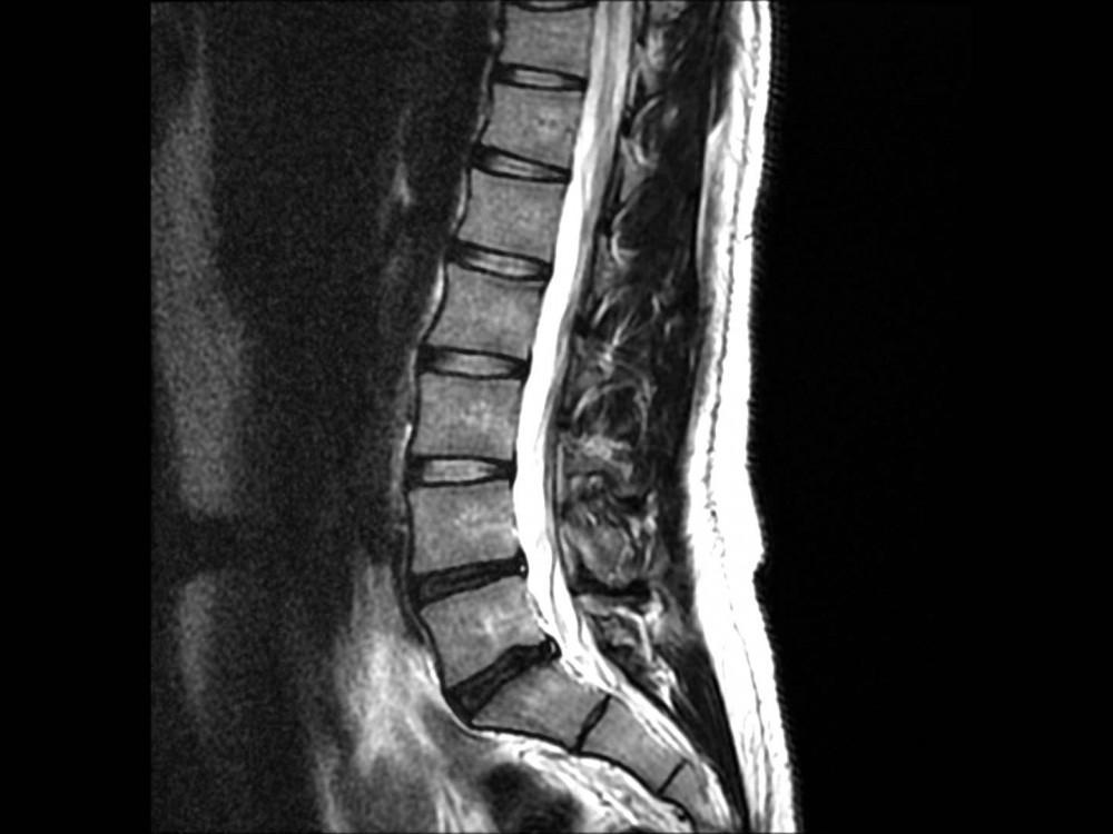
Magnetic resonance imaging (MRI) is a type of scan that uses strong magnetic fields and radio waves to produce detailed images of the inside of the body. This type of imaging gives healthcare professionals a snapshot of your anatomy. Different tissues can all be seen at the same time (bone, muscle, nerve, ligament, etc.). Different structures and fluids are all different shades of light and dark, depending on the type of sequences being used. MRI is usually the investigation of choice, but only for certain indications.
Traditional scans are done in a lying position, and take approximately 30 minutes to perform a single body part. The best images are always taken in a machine where you are lying in a tube-like machine. However, there are options for open MRI scans which are usually preferred by those with claustrophobia. Unfortunately, the open scanners have reduced image quality.
So why shouldn't I get an MRI for my back pain?

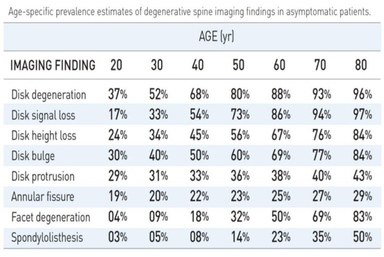
It is common to think that if you have pain, then an MRI scan will find it. It makes sense to think that imaging can find the source of the pain with 100% accuracy. Unfortunately, it is much more complicated than this. See the above table for example, taken from a study which can be seen here.
What this table shows is the NORMAL age-related changes in those with no back pain at all. The left of the table shows a lot of different structural changes which can be seen on an MRI scan. The following columns show how common these changes are depending on age (across the top). What this shows is how normal a lot of different MRI findings are. For example, when you are 40 years old, disc bulges are present in 50% of people without any back pain at all.
This makes a lot of clinical decision-making difficult. If we know that a certain finding or structural change is common in your age group in people without any pain, then can we really call this a problem? If a scan is performed without any context of symptoms or pain, then the scan does not factor into decision-making. An MRI is only useful for the right person and at the right time.
When do I need an MRI scan for my low back pain?
MRI scans are useful to have early depending on the circumstances. So when are these?
- Worsening and progressive symptoms.
- Failure to respond to first-line treatment/management.
- Associated leg symptoms causing marked changes (muscle weakness, vast sensation changes, balance disturbances).
- Less common presenting symptoms (changes in sensation around the groin, or loss of control when going to the toilet).
- When there is suspicion of more sinister problems.
How do I get a lumbar spine MRI?
This should be only be requested by an appropriately qualified healthcare professional. You should have a thorough assessment first to ensure any findings on an MRI scan can be matched against your signs and symptoms. Without this, any scan findings become hard to interpret. Ideally the referring healthcare professional has training in understanding the images, and also has the appropriate knowledge on how to use the findings to help you.
Ideally, the person referring you will also have access to view the images. This ensures that your presenting low back pain can be fully matched against the scan findings. If this doesn’t happen, this may lead down to a path of treating something which doesn’t help your problem. Relying on the report is not best practice, and leaves all of your image interpretation down to the radiologist alone. Although the reporting radiologist’s report is highly valuable, it is the referring healthcare professional who knows the patient and their context the best.
The referring clinician will most commonly be a doctor/consultant who specialises in spinal conditions, or a healthcare professional such as a physiotherapist who has undergone extensive training and can treat or refer as appropriate.
Where can I find more information?
Some physiotherapists are trained to refer and treat MRI scan findings accordingly. These physiotherapists are required to be in a setting which can refer for imaging, and have the contact network to refer onwards as necessary.
Bradley Rugg is a Non-Medical Referrer and has received extensive training in spinal conditions, and using investigations such as MRI scans to help the management of those with low back pain. Any referral will only be performed after an assessment, and is deemed necessary.

